Una nuova era per la diagnostica delle malattie della retina - AngioOCT
 Il ruolo della fluorangiografia retinica pur se in calo negli ultimi anni, è sempre preponderante. In molte malattie come la degenerazione maculare senile, la retinopatia diabetica, le occlusioni vascolari, la fluorangiografia permette di definire con precisione gli aspetti anatomo-fisiologici della malattia,dallo stadio iniziale a quello più avanzato. E questo ruolo assume una connotazione ancora più rilevante se vengono associate l'angiografia con verde indocianina o l'OCT.
La fluorangiografia è ritenuta dalle varie linee guida l'esame prioritario per confermare la diagnosi di degenerazione maculare senile nei casi sospetti, per una classificazione clinica e per iniziare una terapia corretta. Ed ancora nel seguire l'andamento della cura o, qualora questa non fosse efficace, per valutare le altre possibilità terapeutiche. Anche se questo esame nei decenni si è dimostrato sicuro e con scarsi effetti collaterali esistono sempre delle remore più o meno tacite soprattutto in casi di generiche allergie, tanto è che esistono considerazioni medico legali che consigliano la presenza o la vicinanza di un anestesista. In molti centri inoltre si effettuano anche esami ematochimici prima dell'esame. Inoltre in altri casi con accertati precedenti di effetti collaterali come nausea, vomito, ipotensione o in più rari casi di gravidanza, si consiglia prudenza o una visita allergologica preliminare.
Pertanto la possibilità di utilizzare sistemi diversi non legati alla presenza di un contrasto può assumere una connotazione di rilevante importanza .
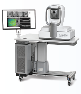
Da qui grazie ai notevoli progressi tecnologici in tale campo l'idea che per effettuare una angiografia si possa utilizzare l'OCT o meglio OCT dedicati di ultimissima generazione con tecnologia 3D. Alcuni svantaggi sono presenti come la visualizzazione limitata alla regione centrale e le difficoltà di esaminare la periferia, la necessità di una ottima collaborazione ed infine la mancata evidenzia del leakage . Un Combo OCT pertanto dotato di OCT tradizionale con modulo per angiografia (angioOct).
E con la attuale tecnologia è già possibile differenziare i vari plessi vascolari, quello superficiale da quello profondo, da quello coroideale e tra breve verificarne il flusso. Gli ambiti di ricerca sono enormi. Per esempio un ridotto flusso coroideale può essere uno dei movens patogenetici per lo sviluppo della degenerazione maculare senile ?
L'importanza dell'Oct oggi è già pienamente consolidata. In questi ultimi 20 anni sono stati fatti miglioramenti eclatanti sulla velocità dello scanning, sulla sensitività e risoluzione. Gli ultimi OCT si basano su uno spettrometro (OCT spectral-domain-SD) o su un laser che varia velocemente (swept-source) con immagini di qualità ottimale e possibilità del 3D.
In conclusione possiamo affermare che al momento attuale la fluorangiografia rimane un esame fondamentale nel processo diagnostico di una lesione neovascolare in corso di degenerazione maculare, nella retinopatia diabetica, nelle trombosi venose e in altre malattie retiniche, sia per visualizzare la regione maculare che la periferia. Tali patologie sin da ora possono pertanto essere diagnosticate e seguite nella loro evoluzione dalla angiografia senza mezzo di contrasto associata al OCT.
Long-Term Outcomes in Eyes Receiving Fixed-Interval Dosing of Anti-VEGF Agents for Wet AMD
The purpose of this single-practice, retrospective chart review was to report on long-term visual outcomes in patients receiving continuous fixed-interval dosing of anti-vascular endothelial growth factor treatment in wet age-related macular degeneration.
It consisted of 109 eyes with exudative AMD receiving continuous fixed-interval dosing (every four to eight weeks) of anti-VEGF therapy (ranibizumab, bevacizumab or aflibercept) for at least five years. Eyes were excluded if they averaged fewer than 6.5 injections per year. Snellen visual acuity was recorded at baseline and all subsequent injections and changes from baseline were calculated at yearly intervals. The primary outcome measure was mean change in letter score at five, six and seven years; secondary outcomes included the percentage of patients with 20/40 vision or better at seven years and the mean change in letter score at each yearly time point based on baseline visual grouping (20/40 or better, 20/50 to 20/100, 20/200 or worse).
A total of 44 patients with seven years of continuous treatment, 75 patients with six years of treatment and 109 patients with five years of continuous treatment were identified. Mean change in letter score at year five was +14.0 letters (p=3.9 × 10⁻⁹), +12.2 letters at six years (p=1.5 × 10⁻⁷) and +12.1 letters at seven years (p=3.8 × 10⁻⁵). It was reported that driving vision (20/40 or better) was achieved in 43.2% of treated eyes. Subanalysis revealed that the greatest visual gains at five and seven years were seen in those patients with baseline visual acuity worse than 20/200 (+24.5 and +25.5 letters), followed by those with 20/50 to 20/100 vision (+6.7 and +6.9 letters), and finally those with 20/20 to 20/40 (+3.7 and +3.4 letters). Patients received an average of 10.5 injections per year.
Continuous fixed-interval dosing of anti-VEGF therapy in patients with exudative AMD results in favorable long-term preservation out to seven years, with vision stabilizing or improving in 93.2% of eyes. Additionally, 43.2% of patients maintained driving vision in the treatment eye at seven years compared with 10.1% at baseline. Our data suggest better outcomes with continuous therapy over published results with sporadic, as-needed therapy.
Source: Peden MC, Suñer IJ, Hammer ME, Grizzard WS. Long-term outcomes in eyes receiving fixed-interval dosing of anti-vascular endothelial growth factor agents for wet age-related macular degeneration. Ophthalmology. 2015;122(4):803–808.
Treatment of Wet AMD With Ranibizumab + Ketorolac Eye Drops or PDT
To evaluate whether ketorolac eye drops plus intravitreal ranibizumab or verteporfin photodynamic therapy plus intravitreal ranibizumab provides additional benefit over intravitreal ranibizumab monotherapy for treatment of choroidal neovascularization in age-related macular degeneration, the authors of this prospective, randomized, pilot study examined 75 patients with naïve CNV.
They randomized patients 1:1:1 into three groups: ranibizumab monotherapy (RM); ranibizumab plus ketorolac; or ranibizumab plus loading-phase reduced-fluence verteporfin PDT (RV) groups.
At 12 months, the authors observed that all groups showed significant improvement in both best-corrected visual acuity and central retinal thickness. They noted that the mean BCVA change from baseline to 12 months was –0.14 ± 0.52 logMAR (20/73 ± 20/29) in the RM group, –0.25 ± 0.60 logMAR (20/46 ± 20/27) in the ranibizumab plus ketorolac group and -–0.10 ± 0.30 (20/97 ± 20/40) logMAR in the RV group. They also reported that mean central retinal thickness change from baseline to 12 months was –125 ± 15 µm in the RM group, –141 ± 21 µm in the ranibizumab plus ketorolac group and –130 ± 15 µm in the RV group. Moreover, both ranibizumab plus ketorolac and RV groups required fewer intravitreal ranibizumab treatments than RM.
Compared with RM and ranibizumab plus verteporfin PDT, the combination of 0.45% ketorolac eye drops t.i.d. and ranibizumab in patients with CNV provided superior BCVA and CRT outcomes. Both combination regimens required fewer intravitreal ranibizumab injections than RM during the 12-month follow-up period.
Source: Semeraro F, Russo A, Delcassi L, et al. Treatment of exudative age-related macular degeneration with ranibizumab combined with ketorolac eyedrops or photodynamic therapy. Retina. 2015;March 16. [Epub ahead of print]. DOI: 10.1097/IAE.0000000000000525.
Scotopic and Photopic Microperimetry in Patients With Reticular Drusen and AMD
Clinical observations suggest that patients with age-related macular degeneration have vision problems, particularly in dim light conditions. Previous studies on structural-functional analysis in patients with AMD with reticular drusen have focused on photopic sensitivity testing but have not specifically assessed scotopic function.
In an effort to evaluate retinal function by scotopic and photopic microperimetry in patients with AMD and a well-demarcated area of reticular drusen, investigators performed this prospective case series in a referral center of 22 eyes from 18 patients (mean age, 74.7 years; range, 62 to 87 years) between June 1, 2014 and October 31, 2014. They used combined confocal scanning laser ophthalmoscopy and spectral-domain optical coherence tomography imaging to identify retinal areas with reticular drusen (category one) and no visible pathologic alterations (category two) in each eye. They also performed scotopic and photopic microperimetry (MP-1S; Nidek Technologies) using a grid with 56 stimulus points. The mean outcome measure was a comparison of mean threshold sensitivities for each category for scotopic and photopic microperimetry.
In all eyes, areas of category one showed a relative and sharply demarcated reduction of scotopic threshold values compared with areas of category two, but only less pronounced differences were seen for photopic testing. According to the investigators, statistical analysis in the 18 eyes in which the 1.0–log unit neutral density filter was applied revealed a difference of scotopic threshold values in areas of category one (mean, 13.5 dB [95% confidence interval, 12.1 to 15.0]) vs. category two (mean, 18.3 dB; [95% CI, 17.4 to 19.3] (p≤0.001). For photopic testing, the mean threshold values were 16.8 dB (95% CI, 15.5 to 18.2) in category one and 18.4 dB (95% CI, 17.1 to 19.6) in category two (p=0.03).
The results of this study suggest that rod function is more severely affected than cone function in retinal areas with reticular drusen. This differential structural-functional correlation underscores the functional relevance of reticular drusen in patients with AMD.
Source: Steinberg JS, Fitzke FW, Fimmers R, et al. Scotopic and photopic microperimetry in patients with reticular drusen and age-related macular degeneration. JAMA Ophthalmol. 2015;March 26. [Epub ahead of print]. DOI: 10.1001/jamaophthalmol.2015.0477.
Half-Dose PDT Combined with Bevacizumab for PCV
To evaluate the efficacy of half-dose photodynamic therapy combined with intravitreal bevacizumab for polypoidal choroidal vasculopathy.
For this retrospective comparative study, data from 57 patients (63 eyes) with at least 12 months of follow-up were reviewed. Full-dose or half-dose PDT combined with a single intravitreal bevacizumab treatment was performed according to the time period. From three months after the initial combination treatment, retreatment was performed mainly using anti-vascular endothelial growth factor injections on an as-needed basis.
Consecutively, 33 eyes were treated with full-dose PDT/intravitreal bevacizumab and 30 eyes with half-dose PDT/intravitreal bevacizumab. At month three, half-dose PDT/intravitreal bevacizumab induced negligible damage to the physiologic choroid but was inferior to full-dose PDT/intravitreal bevacizumab in achieving complete polyp closure (43.3% vs. 72.7%, p=0.018) and improving mean best-corrected visual acuity (20/66 vs. 20/43, p=0.020). At month 12, the half-dose group achieved comparable visual improvement (20/51 vs. 20/40, p=0.254) but required more additional injections (a mean of 2.80 vs. 1.03, p=0.004).
Despite inferior efficacy in inducing polyp closure, half-dose PDT/intravitreal bevacizumab followed by additional injections showed promising visual outcomes while avoiding damage to the physiologic choroid. Further long-term study is needed to evaluate the efficacy of half-dose PDT plus anti-VEGF for PCV.
Source: Lee JH, Lee WK. Half-dose photodynamic therapy combined with bevacizumab for polypoidal choroidal vasculopathy. Retina. 2015;March 12. [Epub ahead of print]. DOI: 10.1097/IAE.0000000000000498.
Intravitreal Aflibercept in the Treatment of Myopic CNV
Researchers seeking to evaluate intravitreal aflibercept 2 mg in patients with myopic choroidal neovascularization conducted this international, Phase III, multicenter, randomized, double-masked, sham-controlled study.
They included patients aged ≥18 years with high myopia (≤–6.0D or axial length of ≥26.5 mm), active myopic CNV and best-corrected visual acuity of 73 to 35 Early Treatment Diabetic Retinopathy Study letters in the study eye. They randomized patients 3:1 to intravitreal aflibercept or sham. Patients in the intravitreal aflibercept arm received one injection at baseline. The study researchers administered additional injections in case of CNV persistence or recurrence at monthly visits through week 44. In the sham arm, patients received sham injections through week 20. At week 24, after assessment of the primary efficacy endpoint, sham patients received a mandatory intravitreal aflibercept injection followed by intravitreal aflibercept (if disease persisted/recurred) or sham injection every four weeks. The main outcome measure was the mean change in BCVA from baseline to week 24.
Acorrding to the researchers, 122 patients were randomized to intravitreal aflibercept (n=91) or sham (n=31). They reported that baseline demographics were similar across groups. At week 24, patients in the intravitreal aflibercept and sham groups gained 12.1 and lost 2 letters, respectively (p<0.0001).
By week 48, patients in the intravitreal aflibercept and sham/intravitreal aflibercept groups gained 13.5 and 3.9 letters. Patients in the intravitreal aflibercept group received two injections (median) in the first study quarter (weeks zero through eight). Median number of injections in quarters two to four was zero. Patients in the “sham/intravitreal aflibercept” group received two and one (median) intravitreal aflibercept injections in quarters three and four. Central retinal thickness improved in parallel with visual gains. Incidence of ocular adverse events was similar in both groups through week 48 (37.4% vs. 38.7); most were assessed by researchers as mild. No deaths occurred.
To conclude, intravitreal aflibercept 2 mg was effective for treatment of myopic CNV with clinically important visual and anatomic benefits achieved with a limited number of injections given in the first eight weeks of treatment. No new safety concerns occurred with treatment. Intravitreal aflibercept should be considered as a treatment option for myopic CNV.
Source: Ikuno Y, Ohno-Matsui K, Wong TY, et al; on behalf of the MYRROR Investigators. Intravitreal aflibercept in patients with myopic choroidal neovascularization: the MYRROR Study. Ophthalmology. 2015;March 4. [Epub ahead of print]. DOI: http://dx.doi.org/10.1016/j.ophtha.2015.01.025.
Regional Choroid Thickness After Reduced-Fluence PDT for Chronic CSC
The Japanese authors of this prospective, consecutive, interventional case series evaluated macular choroidal thickness after reduced-fluence photodynamic therapy for chronic central serous chorioretinopathy.
They treated 22 eyes with chronic CSC with reduced-fluence PDT and examined macular choroidal thickness using spectral-domain optical coherence tomography with a 3-D radial scan protocol in the choroidal mode before and one, three and six months after the treatment. They also compared the mean choroidal thickness in the Early Treatment Diabetic Retinopathy Study grid (center, inner circle and outer circle) between before and after therapy, as well as between treated eyes and 54 volunteer normal eyes.
Chronic CSC eyes showed significantly thicker choroids in the macular area compared with normal controls (p<0.0001), the authors observed. After the single treatment session, they noted that subretinal fluid resolved completely in all eyes, and there were no recurrences during the study period. Additionally, choroidal thickness within the center area and inner circle showed a significant reduction at all time points after treatment (p<0.05). The choroidal thickness in the outer circle showed a statistically significant reduction at one and three months but not at six months. After treatment, the choroidal thickness reduced to the normal values at the center and inner circle, but was still significantly thicker in the outer circle (p<0.01).
The study authors concluded that chronic CSC eyes showed significantly thicker choroids in the macular area. After reduced-fluence PDT, macular choroidal thickness became thinner within six months of treatment.
Source: Manabe S, Shiragami C, Hirooka K, et al. Change of regional choroid thickness after reduced-fluence photodynamic therapy for chronic central serous chorioretinopathy. Am J Ophthalmol. 2015;159(4):644–651.
Connection Between FAF and Central Visual Function in Chronic CSC
To find possible correlations between the morphologic macular changes revealed by fundus autofluorescence and the functional parameters such as visual acuity and retinal sensitivity in patients with chronic central serous chorioretinopathy, investigators in Italy conducted this prospective, cross-sectional study.
They studied 46 eyes (39 consecutive patients) with chronic CSC using FAF and microperimetry. Retinal sensitivity value maps were exactly superimposed over FAF images and the following microperimetric parameters were applied: central 10-degree visual field; 4-2-1 strategy; 61 stimulation spots; white monochromatic background; stimulation time 200 ms; and stimulation spot size Goldmann III. The investigators also explored a possible relationship between microperimetry and FAF.
They discovered that mean best-corrected visual acuity was 20/32 (median 20/25, range 20/20 to 20/200) and that BCVA was significantly correlated with FAF findings (Mann-Whitney test; p<0.0001). They also found a positive concordance between FAF and microperimetry evaluation (total concordance of 0.720 with a kappa of Cohen of 0.456). They noted that the hypo-autofluorescent areas showed decreased retinal sensitivity, while adjacent areas of increased FAF could be associated to both normal and decreased retinal sensitivity. Furthermore, absolute scotoma, defined as 0 dB retinal sensitivity, corresponded with absence of autofluorescence.
Altered FAF in chronic CSC patients has a functional correlation quantified by microperimetry. This study confirms the impact of FAF changes on retinal sensitivity and their value to reflect the functional impairment in chronic CSC.
Source: Eandi CM, Piccolino FC, Alovisi C, et al. Correlation between fundus autofluorescence and central visual function in chronic central serous chorioretinopathy. Am J Ophthalmol. 2015;159(4):652–658.
Therapies for Macular Edema Associated with CRVO
In an effort to review the available evidence regarding the safety and efficacy of therapies for the treatment of macular edema associated with central retinal vein occlusion, a literature search of the PubMed database was last conducted in March 2014 with no date restrictions but limited to articles published in English. A literature search of the Cochrane Library was also conducted in March 2014 with no date restrictions and without a language limitation. The combined searches yielded 108 citations, of which 20 were deemed clinically relevant for the Ophthalmic Technology Assessment Committee Retina/Vitreous panel to review in full text. Three additional studies were also identified for panel review. The level of evidence of these selected studies was reviewed by the panel methodologist.
There were seven citations representing four clinical trials that provided level I evidence supporting the use of anti-vascular endothelial growth factor pharmacotherapies for macular edema associated with CRVO, including: intravitreal ranibizumab (two); aflibercept (three); and bevacizumab (two). There were three citations representing two studies with level I evidence for intravitreal corticosteroid injection with dexamethasone intravitreal implant (two citations) or triamcinolone (one citation), although cataract and glaucoma were observed in these studies. Level I evidence is available on the limited benefit of macular grid-pattern laser photocoagulation (one citation). Eight other citations reviewed were rated as level II, and four citations were rated as level III. Long-term efficacy results (&#ge;2 years of follow-up) are limited to intravitreal ranibizumab at this time, and few studies have evaluated combination therapy with anti-VEGF and corticosteroid versus monotherapy of either class of drug.
Level I evidence indicates that intravitreal anti-VEGF pharmacotherapy is safe and effective over two years for macular edema associated with CRVO and that delay in treatment is associated with worse visual outcomes. In addition, level I evidence demonstrates short-term efficacy of intravitreal corticosteroid but also an association with a higher frequency of adverse events.
Source: Yeh S, Kim SJ, Ho AC, et al. Therapies for macular edema associated with central retinal vein occlusion: a report by the American Academy of Ophthalmology. Ophthalmology. 2015;122(4):769–778.